What are Gliomas?
Gliomas are most common primary brain tumors . Gliomas are thought to be arising from supportive cells in brain called glial cells.
⏱ Avg. Reading Time: 3 min
Gliomas are most common primary brain tumors . Gliomas are thought to be arising from supportive cells in brain called glial cells.
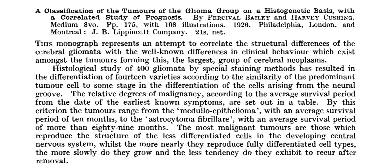
1926: Classification based on cells of origins
Bailey and Cushing in 1926, demonstrated that the cellular structure of a tumor can guide treatment and prognosis.
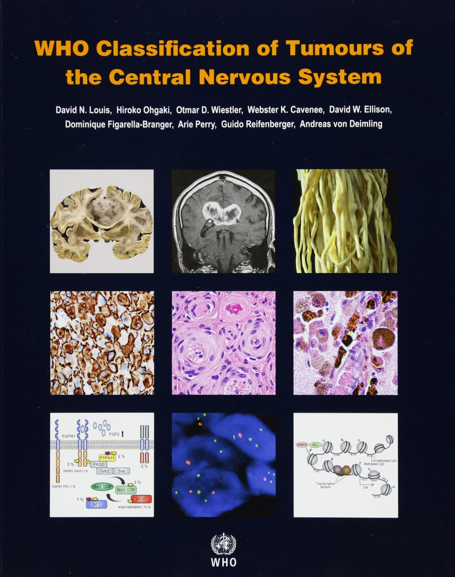
2016 WHO Brain Tumor Classification
2016 WHO Classification which updated the 2007 WHO Classification, considered genetic markers for the first time in the WHO classification. This was to in keep in pace with advances in the genetic and molecular biology. IDH mutation and 1p19q codeletion formed the basis for this classification for gliomas.
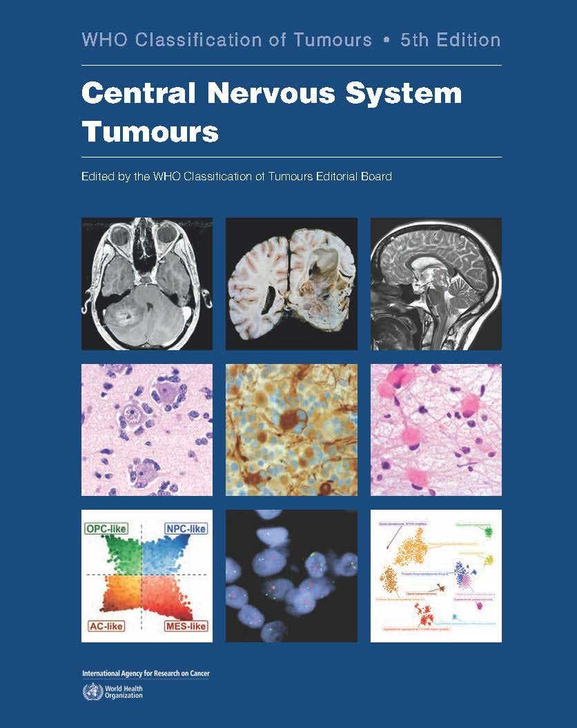
2021 WHO Brain Tumor Classification
Even though the update of previous classification from 2007 took almost a decade this newer classification was published in just 4 years. This can give us some insight into how exponential field of genetics is evolving. cIMPACT updates which were published after 2016 WHO classifications were the basis of this new classification. This classification describes a subset of astrocytomas which may appear low grade on histology but show survival as poor as high grade glioblastomas.
They are called molecular glioblastomas.
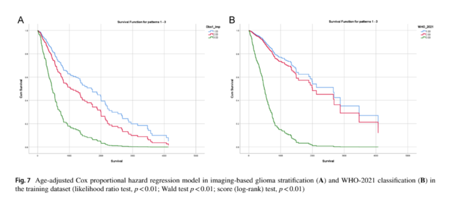
2022: Imaging-based stratification of adult gliomas prognosticates survival and correlates with the 2021 WHO classification
Study done by our group utilised stratified gliomas using a novel flowchart based approach. Imaging-based stratification was statistically significant in predicting survival in both datasets with good inter-observer agreement (age-adjusted Cox-regression, AUC > 0.5, k > 0.6, p < 0.001). IBGS type-I, type-II, and type-III gliomas had good specificity in identifying IDHmut 1p19q-codel oligodendroglioma (training - 97%, validation - 85%); IDHmut 1p19q non-codel astrocytoma (training - 80%, validation - 85.9%); and IDHwt glioblastoma (training - 76.5%, validation- 87.3%) respectively (p-value < 0.01).
2023: GliomaAI
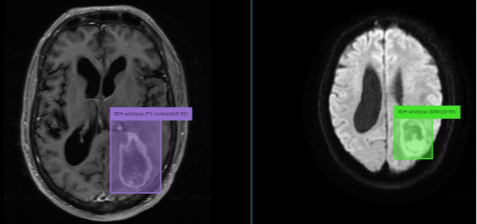
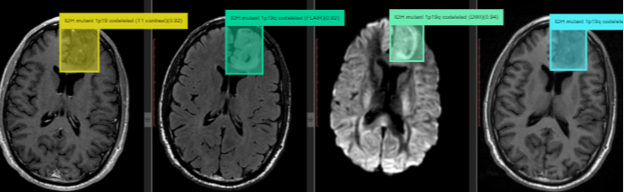
Glioma AI is in making since 2017, since the WHO 2016 update. It has undergone making updates with cIMPACT publications and with recent WHO 2021 classification. It was validated for an year. Novel hierarchial method of deep learning training was used create Glioma AI. It is made by creators of Imaging Based Glioma Stratification (IBGS). Glioma AI was trained on 691 MRI examinations from TCIA (Tumor Cancer Imaging Archieve). The Glioma AI in showed precision of 0.88, recall of 0.8, and F1 score of 0.84 in stratifying gliomas, with an average inference time of 0.07 sec in differentiating adult gliomas into one of three genetic categories, isocitrate dehydrogenase (IDH)-mutant/1p19q-codeleted oligodendroglioma, IDH-mutant/1p19q-intact astrocytoma, and
IDH-wildtype astrocytoma/glioblastoma.
2023: GliomaAI.org, post deployment trial
Readers of the research paper will be able to submit the radiology scans for glioma characterization by Glioma AI (GliomaAI.org). The MRI data would be anonymized in the upload pipeline. The uploading users would get a hybrid diagnosis result after Glioma AI results are checked by neuroradiologists. We would then ask users to submit the genetic results and other feedback. This trial would run for the next 6 months. The cross-table analysis will be performed by comparing the Glioma AI results with the genetic results provided by users. The accuracy of both GliomaAI and human neuroradiologist reader would also be compared. The results of the cross-table analysis would be published in the future study.
Three public datasets were collected from The Cancer Imaging Archive (TCIA). These datasets contain the MRI data from 12 different institutions, containing total of 691 MRI examinations.
218 cases from the training database were used in the internal validation.
329 were cases were used in the external validation and compared with human experts.
Glioma AI had a precision of 0.88, recall of 0.8, and F1 score of 0.84 in stratifying gliomas, with an average inference time of 0.07 sec in differentiating adult gliomas into one of three genetic categories, isocitrate dehydrogenase (IDH)-mutant/1p19q-codeleted oligodendroglioma, IDH-mutant/1p19q-intact astrocytoma, and IDH-wildtype astrocytoma/glioblastoma.
Cross-table analysis of specificity and sensitivity for IDH mutant/1p19q-codeleted oligodendroglioma in validation dataset.
Cross-table analysis of specificity and sensitivity for IDH mutant/1p19q non-codeleted astrocytoma in validation dataset.
Cross-table analysis of specificity and sensitivity for IDH wildtype (Histological Glioblastoma) in validation dataset.
Cross-table analysis of specificity and sensitivity for IDH wildtype molecular glioblastoma in validation dataset.
WHO 2021 classification
Approximately 100,000 people are diagnosed with gliomas worldwide every year. Gliomas lead to substantial mortality and morbidity. While gliomas were historically classified based on their cells of origin, recent advances in molecular genetics markers have led to the 2021 WHO classification update of diffuse gliomas.
The Gap
Recently published survival trend analysis has shown increased survival trends in affluent countries, with an increasing gap between developing and developed countries [1]. The proposed Glioma AI can decrease this gap by providing timely radio-genomic correlation based on non-invasive MRI imaging. The application of such an artificial intelligence model could have a significant impact on patient management especially when stereotactic biopsy and genetic testing are limited by co-morbidities or financial constraints.
Girardi, F., Matz, M., Stiller, C., You, H., Marcos Gragera, R., Valkov, M. Y., … Lewis, C. (2022). Global survival trends for brain tumors, by histology: analysis of individual records for 556,237 adults diagnosed in 59 countries during 2000–2014 (CONCORD-3). Neuro-Oncology, 25(3), 580–592. https://doi.org/10.1093/neuonc/noac217
The Solution
The novel hierarchical deep learning model (Glioma AI) stratified gliomas into three categories which correlate with WHO 2021 classification. Glioma AI showed good precision in both the validation dataset and the deployment trial. Class precision in identifying gliomas in both validation and deployment datasets was comparable.
Post Publication Feedback
Our study also has integrated a post-publication feedback framework about the proposed novel deep learning model, Glioma AI. This will provide readers a tool to upload the MRI data for getting results from the proposed Glioma AI. To the best of our knowledge, this is the first study that gives readers an opportunity to access the model’s inference along with hybrid results.
WHO 2021 classification
Approximately 100,000 people are diagnosed with gliomas worldwide every year. Gliomas lead to substantial mortality and morbidity. While gliomas were historically classified based on their cells of origin, recent advances in molecular genetics markers have led to the 2021 WHO classification update of diffuse gliomas.
The Gap
Recently published survival trend analysis has shown increased survival trends in affluent countries, with an increasing gap between developing and developed countries [1]. The proposed Glioma AI can decrease this gap by providing timely radio-genomic correlation based on non-invasive MRI imaging. The application of such an artificial intelligence model could have a significant impact on patient management especially when stereotactic biopsy and genetic testing are limited by co-morbidities or financial constraints.
Girardi, F., Matz, M., Stiller, C., You, H., Marcos Gragera, R., Valkov, M. Y., … Lewis, C. (2022). Global survival trends for brain tumors, by histology: analysis of individual records for 556,237 adults diagnosed in 59 countries during 2000–2014 (CONCORD-3). Neuro-Oncology, 25(3), 580–592. https://doi.org/10.1093/neuonc/noac217
The Solution
The novel hierarchical deep learning model (Glioma AI) stratified gliomas into three categories which correlate with WHO 2021 classification. Glioma AI showed good precision in both the validation dataset and the deployment trial. Class precision in identifying gliomas in both validation and deployment datasets was comparable.
Post Publication Feedback
Our study also has integrated a post-publication feedback framework about the proposed novel deep learning model, Glioma AI. This will provide readers a tool to upload the MRI data for getting results from the proposed Glioma AI. To the best of our knowledge, this is the first study that gives readers an opportunity to access the model’s inference along with hybrid results.
With the popularization of glioma diagnosis methods, its rapidly increasing incidence rate began to decelerate and has continued to decline since 1987. However, the glioblastoma incidence trend was different, with a significant increase of 0.2% per year after 1992. Thus, changes in the use of cancer screening tests are not solely responsible for the incidence trends.
Ref: Lin D, Wang M, Chen Y, Gong J, Chen L, Shi X, Lan F, Chen Z, Xiong T, Sun H, Wan S. Trends in intracranial glioma incidence and mortality in the United States, 1975-2018. Frontiers in Oncology. 2021 Nov 1;11:748061.
Vigneswaran K, Neill S, Hadjipanayis CG. Beyond the World Health Organization grading of infiltrating gliomas: advances in the molecular genetics of glioma classification. Annals of translational medicine. 2015 May;3(7).
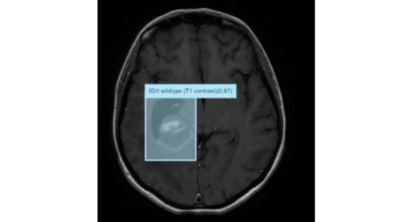
Glioma near right lateral ventricle
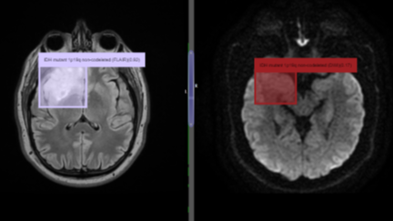
Right temporal lobe lesion. Usually they are high grade IDH wild. However in this case it demonstrates Glioma AI does not reply only on the distribution of tumor.
How deep learning really works it's still a mystery. Even though gross specimens were not used to train the Glioma AI. It has picked up lesions in the gross brain specimens as well.